01 — The problem
Hours, not days
Acute brain slices are the workhorse preparation for studying neural circuits, and optogenetics has made them more useful still. But a slice in a conventional chamber degrades within hours, which rules out any protocol that needs to observe the same tissue over a long window — repeated stimulation, slow plasticity, anything where the interesting change takes longer than the tissue lasts.
02 — The approach
A fluidics problem with optical constraints
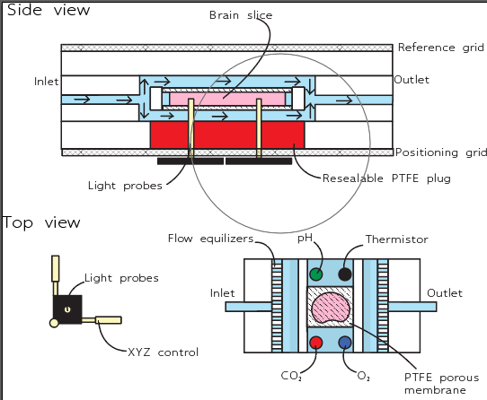
The chamber perfuses the slice continuously rather than bathing it, keeping oxygen and nutrient delivery steady across the tissue instead of letting gradients build. Designing it is a fluidics problem constrained by an optical one: the slice has to be accessible to light and to a probe while the flow field stays uniform over it.
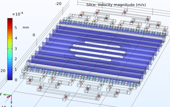
Modelling the velocity field is how that uniformity gets checked before anything is machined — the flow equalisers exist because the first geometries put a gradient straight across the tissue.
03 — Where it stands
Two days down to two hours
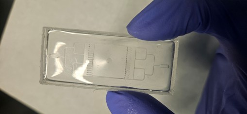
I lead the device side of the collaboration. The first version worked but took two full days to build, which made iteration impossibly slow. Reworking the microfabrication process brought that down to two hours, and the design has gone through many more revisions since as a direct result.
The platform is being brought to full functionality now: CNC machining of the chamber body, probe integration for stimulation and recording, and cleanroom fabrication of the microfluidic layer. Each of the three has to work with the other two, so the current phase is as much integration as it is fabrication.
04 — What I took from it
Iteration speed was the real constraint
The build-time result mattered more than it sounds. Cutting a two-day process to two hours did not improve the device directly; it improved the rate at which the device could be wrong and get fixed. On a project with three interacting subsystems, iteration speed turned out to be the real constraint.